Improving breast cancer treatment in northern Alberta
Surgeons at the Misericordia Community Hospital use leading-edge imaging to give patients better health and cosmetic outcomes

October 25, 2021
By Marguerite Watson, Senior Communications Advisor, Covenant Health
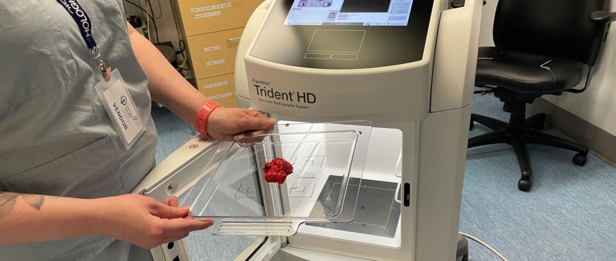
Thanks to the use of a high-definition imaging machine, Heidi Hadubiak felt she could breathe easier after undergoing a lumpectomy at the Misericordia Community Hospital earlier this year. The machine enabled oncoplastic and reconstructive breast surgeon Dr. Lashan Peiris to check in real time, while still in the operating room, that he had successfully removed the cancerous cells from her breast.
“The peace of mind of knowing that my body was cancer-free at that moment was priceless,” says Heidi, 43.
The high-definition specimen radiography system, developed by Faxitron, was purchased for breast cancer surgery at the Misericordia with a $150,000 gift from an anonymous donor to the Covenant Foundation. The purchase came on the heels of recruiting Dr. Nikoo Rajaee, who had used the machine in Ontario, to the Misericordia’s oncoplastic program last year.
More breast cancer surgeries — about 1,500 every year — are performed at the Misericordia than at any other hospital in northern Alberta. Lashan, Nikoo and their colleagues use the Faxitron in the operating room during lumpectomies for both breast cancer and precancerous changes in the breast, and for patients who’ve had chemotherapy, to quickly assess the breast tissue. Within about 20 seconds, they can see the tumour in the specimen and the area, or surgical margin, around it.
“We want to take the cancer out and have nice healthy breast tissue all the way around the cancer,” says Lashan. “If there’s cancer at the margin, that increases the risk of a reoccurrence in the breast.”
Before having the Faxitron machine, breast surgeons at the Misericordia relied on pathologists to come to the operating room to slice the specimen and “eyeball it,” says Lashan. With the Faxitron, surgeons can detect additional lumps in the tissue that aren’t visible to the naked eye or can’t be felt, such as tiny spots of calcification.
“It makes breast surgery much more efficient and efficacious,” says Nikoo. “I’ve noticed that I don’t have to go back for as many margins as I did before.”
In Heidi’s case, the imaging revealed cancerous specks at the margin, prompting Lashan to remove an extra piece of tissue. That meant Heidi didn’t have to have a second surgery in that area of her breast at a later date, which would have created more anxiety for her and her family and prolonged her recovery.
“Being able to recover and have that leg of the journey done is really important from a quality-of-life perspective,” says Heidi.
By reducing the need for repeat surgeries, use of the machine has lessened the strain on the hospital’s operating room space and resources, says Carol Price, program manager, operating room and Faxitron project lead. And access to quick imaging has shortened surgery times, leading to patients spending less time under anesthesia and decreasing risks.
“It has improved workflow efficiencies and led to reduced costs as well,” says Carol.
Research also suggests that patients get a better cosmetic outcome as a result of use of the machine. A hospital in the United Kingdom has reported that the weight of their lumpectomy specimens decreased after surgeons began using the Faxitron, says Lashan.
“They were being more conservative with their lumpectomies. What that means is that they were actually giving patients a better cosmetic outcome because obviously the more breast tissue you take out, the worse the cosmetic outcome. If we can reduce some breast weight generally without compromising the cancer surgery, it contributes to the patients’ long-term quality of life as well. The indirect and direct effects of this machine are huge.”